California Institute of Technology researchers have developed Single Shot Microscopy (SSM), an advanced imaging technique that captures high-resolution images of molecules in nanoseconds. SSM offers benefits like speed, live-cell imaging, and clarity, and has the potential to revolutionize fields like biomedical research, materials science, and clinical diagnostics.
Copyright infringement not intended
Picture Courtesy: THE HINDU
Researchers at the California Institute of Technology have developed Single Shot Microscopy (SSM), an advanced imaging technique that captures high-resolution images in a single exposure.
Most traditional advanced microscopes either "scan" an area point by point or collect multiple "frames" over time to build up a full image.
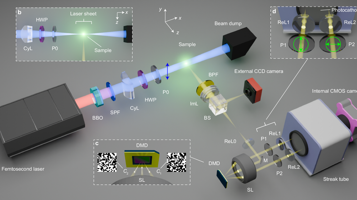
Single Shot Microscope uses a combination of technologies:
Speed => It can "freeze" ultra-fast molecular and cellular dynamics. For example, scientists can now study how proteins fold or how viruses enter cells in real-time.
Live-Cell Imaging => Reduces the total light exposure and heat damage to live cells, which allows to study living cells over extended periods without harming them.
Clarity => By capturing everything in one instant, SSM completely eliminates motion artifacts and blur, show details that would be lost with slower methods.
Biomedical Research => Track drug delivery mechanisms in real-time within cells, seeing exactly where the drug goes and how it interacts, which will help in designing more effective medicines.
Materials Science => Observe rapid crystallization processes or how materials deform and break under sudden stress, which will help to develop stronger, more durable materials for everything from airplane parts to building structures.
Clinical Diagnostics => Revolutionize clinical diagnostics, faster, more accurate pathogen identification (like quickly finding out what bacteria is causing an infection) or detecting cellular abnormalities (like early signs of cancer) almost instantly.
Must Read Articles:
India Waives Local Clinical Trials for Approved Drugs
Source: THE HINDU
|
PRACTICE QUESTION Q. Consider the following statements in the context of the "Single-Shot Microscopy": 1. It captures an entire 3D volume or data set from a sample in a single exposure. 2. It primarily relies on mechanical scanning to build high-resolution images. 3. It can significantly reduce phototoxicity and photobleaching in live-cell imaging. Which of the statements given above are correct? A) 1 only B) 2 and 3 only C) 1 and 3 only D) 1, 2 and 3 Answer: C Explanation: Statement 1 is correct: Single-shot microscopy's defining feature is its ability to capture data instantaneously, which reduces imaging time. Statement 2 is incorrect: Single-shot microscopy avoids mechanical scanning; a characteristic of traditional methods. It uses advanced optical and computational techniques for instantaneous data acquisition. Statement 3 is correct: By reducing the total light exposure time, single-shot microscopy minimizes damage to sensitive live samples, thereby reducing phototoxicity (cell damage due to light) and photobleaching (loss of fluorescence due to light exposure). |
Single-shot microscopy is a technique that captures all the necessary data for an image, including 3D information, in a single, instantaneous exposure, unlike traditional methods that build images sequentially.
Traditional microscopes often rely on scanning or multiple exposures to build an image, whereas single-shot microscopy captures the entire image or volumetric data at once, significantly speeding up the process.
Live cells are dynamic, and rapid imaging prevents motion blur, reduces light exposure (minimizing damage or "phototoxicity"), and allows scientists to observe fast biological processes in real-time.


© 2026 iasgyan. All right reserved