Description
Disclaimer: Copyright infringement not intended.
Context
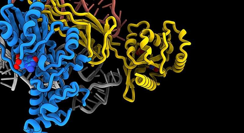
Using electron microscopy, researchers have managed to produce a 3D model of a part of the human cell, the ribosome, which is no more than 30 nanometers in diameter.
Details
Introduction to Ribosomes
- Ribosomes are essential cellular organelles responsible for protein synthesis in all living organisms.
- They are complex macromolecular machines composed of ribosomal RNA (rRNA) and proteins.
- Ribosomes play a central role in translating genetic information from mRNA into functional proteins, making them indispensable for cell growth, proliferation, and maintenance of cellular functions.
Structure of Ribosomes:
- Composition: Ribosomes consist of two subunits - a large subunit and a small subunit.
- Eukaryotic Ribosomes: In eukaryotic cells, the ribosome is composed of a 60S large subunit and a 40S small subunit, forming an 80S ribosome.
- Prokaryotic Ribosomes: Prokaryotic ribosomes are smaller, consisting of a 50S large subunit and a 30S small subunit, forming a 70S ribosome.
- Ribosomal RNA (rRNA): rRNA provides the structural scaffold for ribosomes and catalyzes peptide bond formation during protein synthesis.
- Ribosomal Proteins: Proteins associate with rRNA to stabilize the ribosome structure and participate in various functions during protein synthesis.
Function of Ribosomes:
- Protein Synthesis: Ribosomes catalyze the translation of mRNA sequences into polypeptide chains.
- Translation Process: Ribosomes decode the genetic information carried by mRNA in the form of codons (three-nucleotide sequences) and assemble amino acids into a polypeptide chain according to the mRNA sequence.
- Aminoacyl-tRNA Binding Sites: Ribosomes contain three binding sites - the A site (aminoacyl-tRNA site), P site (peptidyl-tRNA site), and E site (exit site) - where tRNA molecules bind and undergo the steps of translation.

Ribosome Synthesis:
- Nucleolar Synthesis: Ribosomes are primarily synthesized in the nucleolus, a subnuclear compartment.
- Transcription of rRNA Genes: rRNA genes are transcribed by RNA polymerase I to produce precursor rRNA transcripts.
- rRNA Processing: Precursor rRNA transcripts undergo extensive processing, including cleavage and modification, to generate mature rRNA molecules.
- Assembly of Ribosomal Subunits: Ribosomal proteins synthesized in the cytoplasm are imported into the nucleus, where they assemble with rRNA to form ribosomal subunits.
Regulation of Ribosome Biogenesis:
- Nutrient and Growth Factor Signaling: Ribosome biogenesis is tightly regulated in response to cellular nutrient and energy status, as well as growth factor signaling pathways.
- Transcriptional Regulation: Transcription of rRNA genes is controlled by specific transcription factors and signaling cascades that respond to cellular signals.
- Post-transcriptional Regulation: Processing and assembly steps of ribosome biogenesis are subject to regulation by various RNA-binding proteins and ribosome assembly factors.
Significance of Ribosomes:
- Cellular Homeostasis: Ribosomes are essential for maintaining cellular homeostasis by ensuring the production of proteins required for cell growth, metabolism, and function.
- Disease Implications: Dysregulation of ribosome biogenesis and function is associated with various human diseases, including cancer, ribosomopathies, and neurodegenerative disorders.
- Antibiotic Targets: Ribosomes are targets for antibiotics that inhibit bacterial protein synthesis, underscoring their importance in antibiotic therapy and drug development.
Introduction to Plant and Animal cell organelles
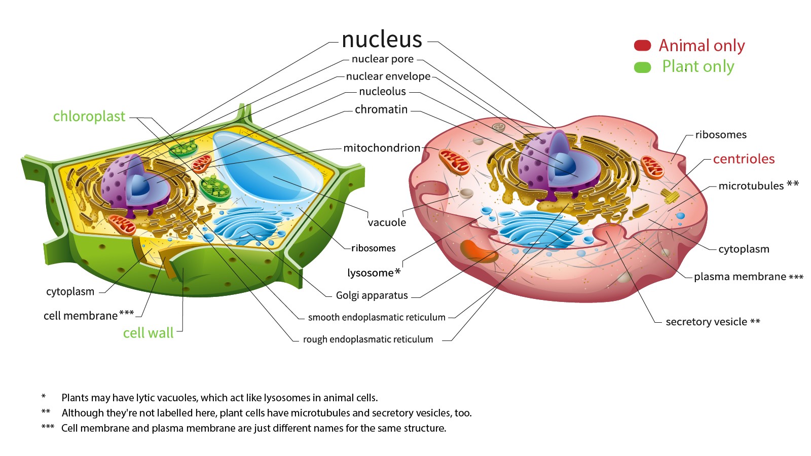
Major organelles found in plant cells:
Nucleus:
- Function: Acts as the control center of the cell, housing the cell's genetic material (DNA) and coordinating cellular activities such as growth, metabolism, and reproduction.
- Structure: Surrounded by a double membrane called the nuclear envelope, containing nuclear pores that regulate the exchange of molecules between the nucleus and the cytoplasm. Contains chromatin (DNA wrapped around proteins), nucleolus (site of ribosome synthesis), and nucleoplasm (fluid within the nucleus).
Chloroplasts:
- Function: Site of photosynthesis, where light energy is converted into chemical energy (glucose) using chlorophyll and other pigments.
- Structure: Surrounded by a double membrane, containing internal membranes called thylakoids organized into grana. Also contains stroma (fluid-filled matrix) where the Calvin cycle occurs.
- Unique Features: Contain their own DNA and ribosomes, allowing them to replicate independently within the cell.
Mitochondria:
- Function: Powerhouse of the cell, generating ATP (adenosine triphosphate) through cellular respiration, which involves the breakdown of glucose and other organic molecules.
- Structure: Surrounded by a double membrane with an inner membrane folded into cristae. Contains matrix (fluid-filled interior) where the Krebs cycle occurs.
- Energy Production: ATP is generated through processes including glycolysis, the Krebs cycle, and oxidative phosphorylation, which occur in different parts of the mitochondria.
Endoplasmic Reticulum (ER):
- Function: Site of protein and lipid synthesis, as well as calcium ion storage and detoxification of drugs and toxins.
- Structure: Consists of rough ER (studded with ribosomes) and smooth ER (lacks ribosomes). Continuous with the nuclear envelope and extends throughout the cytoplasm.
- Rough ER: Involved in protein synthesis and processing, with ribosomes translating mRNA into proteins that are then modified, folded, and transported.
- Smooth ER: Synthesizes lipids, detoxifies harmful substances, and stores calcium ions.
Golgi Apparatus:
- Function: Modifies, sorts, and packages proteins and lipids synthesized in the ER for transport to their destination within the cell or for secretion outside the cell.
- Structure: Consists of flattened membranous sacs called cisternae, arranged in a stack. Has distinct cis and trans faces, with vesicles transporting materials between them.
- Processing Steps: Proteins and lipids from the ER are modified (e.g., glycosylation), sorted, and packaged into vesicles for transport.
Vacuole:
- Function: Plays a role in maintaining turgor pressure, storing water, ions, and nutrients, degrading macromolecules, and serving as a reservoir for secondary metabolites and pigments.
- Structure: Membrane-bound organelle containing a solution called cell sap, surrounded by a single membrane called the tonoplast.
- Central Vacuole in Plant Cells: Occupies a large portion of the cell volume in mature plant cells, exerting pressure against the cell wall to maintain rigidity (turgor pressure).
Cell Wall:
- Function: Provides structural support, protection, and shape to plant cells, preventing cell lysis and allowing for growth and development.
- Structure: Composed primarily of cellulose, a polysaccharide, along with other polysaccharides, proteins, and lignin (in some cells). Lies outside the plasma membrane.
Plasma Membrane:
- Function: Regulates the movement of substances into and out of the cell, maintains cell integrity, and facilitates cell-cell communication.
- Structure: Phospholipid bilayer embedded with proteins and other molecules, with hydrophilic heads facing outward and hydrophobic tails facing inward.
- Selective Permeability: Controls the passage of ions, nutrients, and other molecules through various transport proteins and channels.
Peroxisomes:
- Function: Catalyze various metabolic reactions, including the breakdown of fatty acids, detoxification of hydrogen peroxide, and synthesis of certain lipids.
- Structure: Membrane-bound organelles containing enzymes involved in oxidative reactions.
- Hydrogen Peroxide Detoxification: Contains catalase, an enzyme that converts toxic hydrogen peroxide into water and oxygen.
Ribosomes:
- Function: Site of protein synthesis, where mRNA is translated into polypeptide chains.
- Structure: Composed of ribosomal RNA (rRNA) and proteins, existing as large and small subunits.
- Location: Can be free-floating in the cytoplasm or bound to the endoplasmic reticulum (rough ER), chloroplasts, or mitochondria.
Major organelles found in animal cells:
Nucleus:
- Function: Houses the cell's genetic material (DNA) and serves as the control center of the cell, regulating gene expression and coordinating cellular activities.
- Structure: Surrounded by a double membrane called the nuclear envelope, containing nuclear pores that regulate the exchange of molecules between the nucleus and the cytoplasm. Contains chromatin (DNA wrapped around histone proteins), nucleolus (site of ribosome synthesis), and nucleoplasm (fluid within the nucleus).
Mitochondria:
- Function: Powerhouse of the cell, responsible for generating ATP (adenosine triphosphate) through cellular respiration, which involves the breakdown of glucose and other organic molecules.
- Structure: Surrounded by a double membrane with an inner membrane folded into cristae. Contains matrix (fluid-filled interior) where the Krebs cycle occurs.
- Energy Production: ATP is generated through processes including glycolysis, the Krebs cycle, and oxidative phosphorylation, which occur in different parts of the mitochondria.
Endoplasmic Reticulum (ER):
- Function: Plays a central role in protein synthesis, folding, and transport, as well as lipid synthesis and calcium ion storage.
- Structure: Consists of rough ER (studded with ribosomes) and smooth ER (lacks ribosomes). Continuous with the nuclear envelope and extends throughout the cytoplasm.
- Rough ER: Involved in protein synthesis and processing, with ribosomes translating mRNA into proteins that are then modified, folded, and transported.
- Smooth ER: Synthesizes lipids, detoxifies harmful substances, and stores calcium ions.
Golgi Apparatus:
- Function: Modifies, sorts, and packages proteins and lipids synthesized in the ER for transport to their destination within the cell or for secretion outside the cell.
- Structure: Consists of flattened membranous sacs called cisternae, arranged in a stack. Has distinct cis and trans faces, with vesicles transporting materials between them.
- Processing Steps: Proteins and lipids from the ER are modified (e.g., glycosylation), sorted, and packaged into vesicles for transport.
Lysosomes:
- Function: Serve as the cell's recycling center, containing enzymes that break down and recycle cellular waste, damaged organelles, and foreign substances.
- Structure: Membrane-bound organelles containing hydrolytic enzymes in an acidic environment.
- Autophagy: Lysosomes are involved in autophagy, the process of degrading and recycling cellular components to maintain cellular homeostasis.
Peroxisomes:
- Function: Catalyze various metabolic reactions, including the breakdown of fatty acids, detoxification of hydrogen peroxide, and synthesis of certain lipids.
- Structure: Membrane-bound organelles containing enzymes involved in oxidative reactions.
- Hydrogen Peroxide Detoxification: Contains catalase, an enzyme that converts toxic hydrogen peroxide into water and oxygen.
Ribosomes:
- Function: Site of protein synthesis, where mRNA is translated into polypeptide chains.
- Structure: Composed of ribosomal RNA (rRNA) and proteins, existing as large and small subunits.
- Location: Can be free-floating in the cytoplasm or bound to the endoplasmic reticulum (rough ER).
Plasma Membrane:
- Function: Regulates the movement of substances into and out of the cell, maintains cell integrity, and facilitates cell-cell communication.
- Structure: Phospholipid bilayer embedded with proteins and other molecules, with hydrophilic heads facing outward and hydrophobic tails facing inward.
- Selective Permeability: Controls the passage of ions, nutrients, and other molecules through various transport proteins and channels.
Cytoskeleton:
- Function: Provides structural support, maintains cell shape, facilitates cell movement, and serves as tracks for intracellular transport.
- Components: Consists of microfilaments (actin filaments), microtubules (tubulin polymers), and intermediate filaments, each serving specific functions in cell structure and motility.
Centrioles:
- Function: Play a role in organizing the microtubule cytoskeleton, particularly during cell division (mitosis and meiosis).
- Structure: Composed of microtubule triplets arranged in a cylindrical structure called a centriole.
- Centrosome: Centrioles are found within the centrosome, a region near the nucleus involved in microtubule organization.

Introduction to Electron Microscopy
- Principle: Electron microscopy relies on the wave-like properties of electrons. Electron beams have much shorter wavelengths than visible light, enabling them to resolve smaller details in specimens.
- Types: There are two primary types of electron microscopy: Transmission Electron Microscopy (TEM) and Scanning Electron Microscopy (SEM).
Transmission Electron Microscopy (TEM):
- Principle: In TEM, electrons pass through a thin specimen, and the resulting transmission pattern is used to generate an image.
- Sample Preparation: Specimens are typically fixed, dehydrated, embedded in resin, and thinly sectioned using an ultramicrotome to achieve transparency.
- Imaging Process: Electrons transmitted through the specimen interact with the dense regions, producing contrast variations that are captured by detectors to form an image.
- Resolution: TEM can achieve ultra-high resolution, down to sub-nanometer levels, allowing visualization of cellular organelles, macromolecules, and even individual atoms.
Scanning Electron Microscopy (SEM):
- Principle: In SEM, a focused electron beam scans the surface of a specimen, and secondary electrons emitted from the surface are detected to generate an image.
- Sample Preparation: Specimens are typically fixed, dehydrated, dried, and coated with a thin layer of conductive material (e.g., gold/palladium) to enhance conductivity and imaging quality.
- Imaging Process: The electron beam scans the sample in a raster pattern, and the emitted secondary electrons are collected and used to construct a three-dimensional image of the specimen's surface.
- Resolution: SEM provides high-resolution images with a depth of field that allows for detailed surface topography visualization.
Cryo-Electron Microscopy (Cryo-EM):
- Principle: Cryo-EM is a specialized form of TEM used to study frozen-hydrated specimens at cryogenic temperatures to preserve their native structure.
- Sample Preparation: Specimens are rapidly frozen in a thin layer of vitreous ice, preserving their natural state without the need for chemical fixation or staining.
- Imaging Process: Cryo-EM techniques, such as single-particle analysis and cryo-electron tomography, allow for high-resolution imaging of biomolecules and cellular structures in their native environment.
- Resolution: Recent advances in cryo-EM technology have led to groundbreaking discoveries, including the determination of high-resolution structures of proteins and macromolecular complexes.
Environmental Scanning Electron Microscopy (ESEM):
- Principle: ESEM is a variation of SEM designed to image specimens in their natural, hydrated state and under controlled environmental conditions (e.g., high humidity).
- Sample Preparation: Specimens are typically minimally processed and can be imaged without the need for extensive dehydration or coating.
- Applications: ESEM is useful for studying biological samples, such as live cells, tissues, and hydrated materials, under physiological conditions.
Recent Advances:
- Cryo-Electron Tomography: Enables three-dimensional visualization of cellular structures and macromolecular complexes at near-atomic resolution.
- Correlative Light and Electron Microscopy (CLEM): Integrates fluorescence microscopy with electron microscopy to correlate molecular and cellular dynamics with ultrastructural details.
- Focused Ion Beam Scanning Electron Microscopy (FIB-SEM): Allows for high-resolution imaging of large volumes of tissue through sequential milling and imaging.
Conclusion
Ribosomes are intricate molecular machines crucial for protein synthesis and cellular function. Understanding their structure, function, and regulation provides insights into fundamental cellular processes and has implications for human health and disease. Ongoing research into ribosome biology promises to uncover new therapeutic avenues and deepen our understanding of cellular physiology and pathology.
|
PRACTICE QUESTION
Q. Electron microscopy is a versatile and indispensable tool for studying the structure and function of biological specimens, materials, and nanoscale systems with unparalleled resolution and detail. Discuss. (250 words)
|